If an Electron Microscope Is Used in Which
This type of microscopy is used to observe processes happening on a samples surface. Although the scanning electron microscope is widely used in the scientific field it has some disadvantages also.
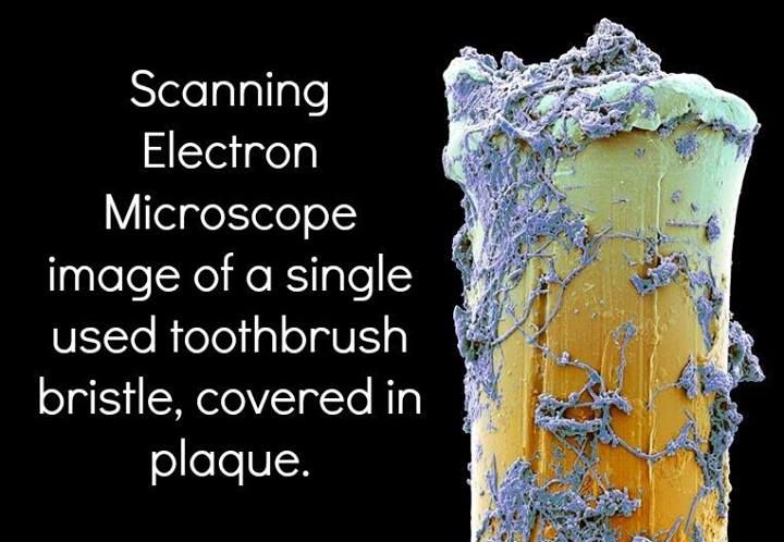
Scanning Electron Microscope Image Of A Single Used Toothbrush Bristle Covered In Plaque Dentist Hygienist Brushing Teeth Toothbrush Bristles Dental Fun
The elastically scattered electrons hit the.

. Reflection Electron Microscope REM With a reflection electron microscope REM an electron beam is on a surface but instead of using the transmission or secondary electrons the beam of elastically scattered electrons is detected. Early History of Electron Microscopy. Perfect preparation makes the difference between trying and achieving between failure and success between results and.
Specimen preparation takes usually takes few days. The invention of the electron microscope by Max Knoll and Ernst Ruska at the Berlin Technische Hochschule in 1931 finally overcame the barrier to higher resolution that had been imposed by the limitations of visible light. Excellent sample preparation is the prerequisite for first-class electron microscopy.
He also aimed at reducing the problems of chromatic aberrations images produced by the. Most electron microscopes used to study biological material can see down to about 10 angstroms--an incredible feat for although this does not make atoms visible it does allow researchers to distinguish individual. In TEM the samples image is formed by the interaction between the.
Electron Microscope Sample Preparation. Electron Microscope Sample Preparation. Overview of Electron Microscopy by Tim Palucka.
Electron microscopes use shaped magnetic. A Transmission Electron Microscope can create a much higher resolution and magnified image than a light microscope because of the shorter wavelength of the electron as compared to photons. Power of the Electron Microscope.
An electron microscope is a microscope that uses a beam of accelerated electrons as a source of illumination. This increased resolution allows us to study ultrastucture of organelles viruses and macromolecules. As the wavelength of an electron can be up to 100000 times shorter than that of visible light photons electron microscopes have a higher resolving power than light microscopes and can reveal the structure of smaller objects.
He used high-resolution power to scan a small raster using a beam of electrons that were focused on the raster. An electron probe is scanning over the surface of the material and these electrons interact with the material. Illuminating source is the Light.
The transmission electron microscope TEM operates on many of the same optical principles as the light microscope. Only solid samples can be analyzed by scanning electron microscopes. However I have gathered some prices from used electron microscopes and included the estimated cost if the microscope were new to give you a sense of the different costs by electron microscope type and model.
Applications of Transmission Electron Microscope TEM TEM is used in a wide variety of fields From Biology Microbiology Nanotechnology forensic studies etc. Scanning electron microscopes are attracted by magnetic fields. To visualize and study cell structures of bacteria viruses and fungi.
Specially prepared materials samples may also be viewed in the TEM. Secondary electrons are emitted from the surface of the specimen and recorded. More detailed story here summary below.
FEI Tecnai G2 F30 Twin. The first Scanning Electron Microscope was initially made by Mafred von Ardenne in 1937 with an aim to surpass the transmission electron Microscope. The height differences in the sample give contrast in the image.
Scanning electron microscope SEM is used to study the topography of materials and has a resolution of 2 nm. To view the shapes and sizes of microbial cell organelles. 1 an electron gun which produces the electron beam and the condenser system which focuses the beam onto the object 2 the image-producing system consisting of the objective lens movable specimen stage and intermediate and projector lenses which focus.
The TEM has the added advantage of greater resolution. Live or Dead specimen may be seen. Condenser Objective and eye piece lenses are made up of glasses.
Be prepared for great results in EM Sample Preparation. Only Dead or Dried specimens are seen. Model Type Used Price Estimated Price New FEI Tecnai F20.
The light microscope and TEM are. The device is extra-large and requires a special room for storage. Skilled and trained operators are required to.
The specimen used in Transmission Electron Microscope should be very thin less than 100 nm thick. Transmission electron microscope TEM type of electron microscope that has three essential systems. To view bacteria flagella and plasmids.
If pushed to the limit electron microscopes can make it possible to view objects as small as the diameter of an atom. Illuminating source is the beam of electrons. Specimen preparation takes usually few minutes to hours.
Some of these applications include.

Compound Light Microscope Electron Microscope Microscope Microscope Parts

Label Microscope Diagram Charts Microscope Polarizing Microscope Anatomy Bones

The 16 Best Science Visualizations Of 2011 Microscopy Science Electron Microscope

Figure 12 These Schematic Illustrations Compare The Components Of Transmission Electron Microsco Microbiology Scanning Electron Microscope Electron Microscope

Different Types Of Microscopes Light Microscope Electron Microscope Scanning Probe Microscope Electron Microscope Confocal Microscopy Optical Microscope
Differences Between Light Microscope And Electron Microscope Electron Microscope Electrons Microscopic

36 Differences Between Light And Electron Microscope Electron Microscope Electrons Scanning Electron Microscope

A Scanning Electron Microscope Image Shows Spherical Particles In Type C Fly Scanning Electron Microscope Images Electron Microscope Images Microscopic Images

Electron Microscope Instrument Electron Microscope Electrons Atomic Structure

Electron Microscope Scanning Electron Microscope Electron Microscope Electrons

Mind Blowing Brain Images From Then And Now Brain Images Electron Microscope Microscopic Photography

Snowflake Images Under An Electron Microscope Electron Microscope Snowflake Images Electron Microscope Images
![]()
Schematic Diagram Of A Transmission Electron Microscope B Scanning Electron Microscope Scanning Electron Microscope Electron Microscope Microscopic Images

Electron Microscope Notes Electron Microscope Electrons Microscopic

How Scanning Electron Microscopes Work Scanning Electron Microscope Electron Microscope Scanning Electron Microscopy

Scanning Electron Microscope Liberal Dictionary Scanning Electron Microscope Electron Microscope Electrons

There Are Two Categories Of Microscopes Based On The Principle On Which Magnificati Scanning Electron Microscope Electron Microscope Electron Microscope Images

Electron Microscope Definition Principle Types Uses Labeled Diagram Electron Microscope Scanning Electron Microscopy Optical Microscope

Scanning Electron Microscope Image Of Sunflower Lower Leaf Surface Note The Electron Microscope Images Scanning Electron Microscope Images Electron Microscope


Comments
Post a Comment